Thursday, July 25, 2013
Cryptorchidism
I have seen plenty of spays and neuters at the clinic. They are a routine, daily occurrence so it is always interesting when something out of the ordinary happens during one of these procedures. The other day there were two neuters scheduled, a cat and a dog. Nothing too exciting or out of the ordinary. However, while the animals were being prepped for surgery it was noticeable that something was not quite right. Both animals had only one testicle. They were both exhibiting cryptorchidism, otherwise known as retained testicles. Cryptorchidism is a condition in which a male's testicles have not descended into the scrotum. During male fetal development, the testes develop high in the abdomen and move from the abdomen down through the inguinal canal and into the scrotum. By the time an animal is at the right age to be neutered, the testes should be fully descended. If they fail to descend, the testicles may remain in the abdomen or inguinal canal. Cryptorchidism can be unilateral in which one testis is retained (like the two neuters scheduled for surgery at the clinic), or it can be bilateral in which both testes are retained. Due to the need for thermal regulation during sperm production, bilateral cryptorchids are sterile while unilateral cryptorchids are still fertile. Cryptorchidism is considered to be an X-linked, autosomal-recessive trait, or in other words a heritable defect, so it is highly advised that cryptorchid animals should not be used for breeding. Surgical removal is the only treatment for cryptorchidism. A cryptorchid surgery is more complicated than a normal neuter because it is more invasive (usually involves opening the abdomen) and the cryptorchid testicle may be difficult to locate. Even animals that are unilateral cryptorchids should have both testicles removed because the retained testicle is prone to testicular torsion and testicular cancer.
Tuesday, July 16, 2013
Forms of External Coaptation
In the time that I have spent at the clinic, I have seen plenty of broken bones. Splints are the "go-to" form of external coaptation that the clinic uses to fixate and immobilize limb injuries. The two most common forms of splints I see used are the Robert Jones Bandage and the Schroeder-Thomas Splint.
The Robert Jones bandage is a common external splint used to immobilize lower-limb injuries. This bandage should not be used on upper-limb fractures/injuries because it cannot properly stabilize such injuries. The Robert Jones bandage offers limb stability, tissue fluid absorption, and protects from trauma. It promotes healing by immobilizing the injured area, thus reducing swelling and providing protection from secondary trauma. The bandage consist of multiple layers of soft, compressible, and absorbent materials such as cotton wrap and conforming bandages. These layers are supported by bandage tape stirrups, plastic splints, or a combination of both on either side of the leg. The entire leg is then wrapped with vet wrap and once finished should keep the leg rigid and immobile.
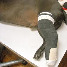
The Schroeder-Thomas splint is another common external splint used to immobilize any fracture below the midfemur of midhumerus. The splint is a traction device or traction splint. This most commonly refers to a splinting device that uses straps attaching over the pelvis, hip, or shoulder as an anchor, metal rods to mimic normal bone stability and limb length, and a sling-like device to apply traction in order to reduce pain and realign the limb. The splint is designed so that bandages and tape are used as slings on the metal frame to position and counteract muscle movement to help align and immobilize parts of the limb.

Although splints are a quick and easy way to set and immobilize fractures, casting is another common form of external coaptation. Casts lie in contact with the skin and are made to conform to the injured area. Plaster and fiberglass can be used for casting, but fiberglass is more commonly used because it is lighter, stronger, and dries quickly. I had the pleasure of seeing a cast applied to a front limb fracture on a dog for the first time the other day. The patient was anesthetized and the fracture was set. Cotton cast padding and a stockingnette were applied to decrease pressure points that could cause skin ulceration. Rolls of casting tape were dunked in warm water and applied up and down the leg. Within minutes the cast was dry and hard as a rock. The cast would be left on for 6-8 weeks in order to allow the fracture to form a callous and heal before being removed.

The Robert Jones bandage is a common external splint used to immobilize lower-limb injuries. This bandage should not be used on upper-limb fractures/injuries because it cannot properly stabilize such injuries. The Robert Jones bandage offers limb stability, tissue fluid absorption, and protects from trauma. It promotes healing by immobilizing the injured area, thus reducing swelling and providing protection from secondary trauma. The bandage consist of multiple layers of soft, compressible, and absorbent materials such as cotton wrap and conforming bandages. These layers are supported by bandage tape stirrups, plastic splints, or a combination of both on either side of the leg. The entire leg is then wrapped with vet wrap and once finished should keep the leg rigid and immobile.
The Schroeder-Thomas splint is another common external splint used to immobilize any fracture below the midfemur of midhumerus. The splint is a traction device or traction splint. This most commonly refers to a splinting device that uses straps attaching over the pelvis, hip, or shoulder as an anchor, metal rods to mimic normal bone stability and limb length, and a sling-like device to apply traction in order to reduce pain and realign the limb. The splint is designed so that bandages and tape are used as slings on the metal frame to position and counteract muscle movement to help align and immobilize parts of the limb.
Although splints are a quick and easy way to set and immobilize fractures, casting is another common form of external coaptation. Casts lie in contact with the skin and are made to conform to the injured area. Plaster and fiberglass can be used for casting, but fiberglass is more commonly used because it is lighter, stronger, and dries quickly. I had the pleasure of seeing a cast applied to a front limb fracture on a dog for the first time the other day. The patient was anesthetized and the fracture was set. Cotton cast padding and a stockingnette were applied to decrease pressure points that could cause skin ulceration. Rolls of casting tape were dunked in warm water and applied up and down the leg. Within minutes the cast was dry and hard as a rock. The cast would be left on for 6-8 weeks in order to allow the fracture to form a callous and heal before being removed.
Thursday, July 11, 2013
Peculiar Poultry
We had the pleasure of treating a rooster at the clinic just the other day. He was a beautiful bird with shiny black feathers and a bright red comb, a show quality bird, but there was one peculiar thing about him. He sat splay legged in his pen with his head tucked under his chest. His neck was so weak and limp that he could barely lift it. The owner explained that it was a perfectly healthy bird and had just started having problems a couple weeks ago, but had gotten progressively worse. The vet examined the rooster and determined that an injury was not the cause of the problem. He concluded that it must be due to a vitamin deficiency. One common deficiency often seen with chickens is riboflavin deficiency. It can be seen rarely in adult birds, but is most commonly seen in young, growing chicks and is often referred to as curled-toe paralysis due to paralysis and disfigurement of the extremities. Sitting splay legged and hock-resting postures are also often seen. The owner was sent home with some riboflavin suspension to give the bird by mouth for 7-10 days, in hopes that the bird would be well and 4-H fair ready within a couple weeks.
Saturday, July 6, 2013
Help! It's stuck!
It seems like it never fails, just when we all think we can close the clinic on time clients walk in at the last minute with animals that need immediate attention and extensive treatment. This always seems to happen on Friday evenings when everyone has plans and is itching to get off work. Just this Friday we had a client call at the last minute to say he was bringing his dog to the clinic because he had a bone stuck in his mouth. We all imagined that the dog had lodged a bone in the back of his mouth and was choking. When the owner arrived with his dog we quickly realized that the dog was in a totally different predicament than we had imagined. The owner had given the dog a stuffed shin bone to chew on. The dog clearly enjoyed his treat a little too much. He had worked so hard to lick the filling from the bone that he somehow worked the bone over his lower jaw and even over his lower canines. The bone was really stuck. The sight was quite hilarious, but the dog was clearly in distress. He clawed frantically at the bone, but it wouldn't budge. The dog was sedated and the veterinarian went to work on trying to remove the bone. He slipped a strand of wire saw underneath the bone and proceeded to saw through. The bone was so tight that the wire saw was starting to cut the dog's skin. The veterinarian went to the clinic's storage room, rummaged around, and came back with a pair of bolt cutters. He placed them at the end of the bone and clamped down. With one cut, the bone snapped off and went flying across the room. The vet laughed and said, "Nothing like an easy and humorous case to end the day."


Subscribe to:
Comments (Atom)