Sunday, August 11, 2013
Organophosphate Toxicity
Recently we had a couple cases at the clinic involving puppies exhibiting signs of sickness and weakness. The pups had been vomiting, having diarrhea, and showing signs of weakness and lack of coordination. At first the signs and symptoms seemed to point towards Parvovirus, a common but very serious disease often seen in puppies. After checking the puppies' records, it was brought to our attention that both were up to date on Parvovirus vaccinations. The pups were tested anyway to be safe, and sure enough both were negative for Parvo. The owners of one puppy said they had applied a topical flea medication and saw symptoms shortly after. The owners of the second puppy had applied Seven dust in order to rid of a flea infestation. The veterinarian dealing with the cases immediately knew what was the cause of the problem. He diagnosed the pups with organophosphate toxicity or in other words, insecticide poisoning. Organophosphate poisoning results from overexposure to organophosphates by inhalation, ingestion, or dermal contact. These compounds are a diverse group of chemicals used in both domestic and industrial settings. Examples include insecticides, herbicides, nerve gases, ophthalmic agents, and anthelmintics. The most common form of organophosphates that pets are exposed to are found in lawn and garden treatments and flea and tick treatments. Organophosphates work by inhibiting acetylcholinesterase, an enzyme critical for nerve function. Inhibition of acetylcholinesterase causes an accumulation of acetylcholine, a neurotransmitter, in the body, which results in nerve and muscle overstimulation. Some noticeable symptoms include fever, vomiting, diarrhea, anorexia, depression, seizures, muscle tremors, hypersalivation, constricted pupils, increased heart rate, lack of coordination, and respiratory failure. Once diagnosed with organophosphate poisoning, the animal needs to be stabilized, decontaminated, and treated. Treatment includes the following, bathing the animal to remove remaining chemical residue, inducing vomiting to flush out the stomach if poison was ingested, administering IV fluids, and administering drugs such as atropine to counteract the effect that organophosphates have on the nervous system. Despite the large variety and widespread use of organophosphates, poisoning can be prevented. Insecticides and other organophosphates should be researched and labels should be followed correctly before using in the home, on the lawn, in the garden, or on pets.
Thursday, August 1, 2013
A Lesson Lived Is A Lesson Learned
One thing I love about working at the clinic is hearing all about the veterinarians wild tales of things that have happened to them during their years of practice. Recently one of the veterinarians was telling a story about drawing blood on a horse for a Coggins test. It was nothing too exciting, just a quick stick and everyone would be on their way. When he went outside the clinic, the owner had not yet unloaded the horse. The veterinarian asked the owner to unload the horse, but he insisted that it would be much easier and quicker just to draw blood from inside the trailer. The veterinarian stepped inside the trailer and slowly approached the horse. The horse, seeing and unfamiliar person coming toward him, began to panic. The vet and owner tried to calm the horse down, but it began to frantically pull against the trailer tie. Suddenly the horse swung its head to one side and snapped the tie. The horse's face collided with the side of the veterinarian's head and knocked him out cold. He had to be dragged from the trailer to avoid being trampled. He woke up outside the trailer with a swollen face, a pounding headache, and a concussion. The vet said he learned the hard way never to get inside a trailer to mess with a horse.
Just this week a client brought in a horse for a Coggins test. The veterinary technician was sent out to draw the blood and I went along to help. It was pouring down rain outside and the owner had not unloaded the horse. The veterinarian's story ran through my head. The owner said that since it was raining we could draw blood from inside the trailer. We stepped into the back of the trailer to get out of the rain, but the veterinary technician suggested that it would be safer to work outside even though it was raining. The owner insisted that we would be fine and proceeded to untie the horse, but just as he loosened the rope the horse spooked and ran backwards. The vet tech and I scurried to get out of the way just as the horse slipped and toppled backwards out of the trailer. The horse regained its footing and luckily wasn't hurt, but the vet tech and I got quite a scare. I think it's safe to say I learned my lesson and will never again get in an unfamiliar horse trailer again.

Just this week a client brought in a horse for a Coggins test. The veterinary technician was sent out to draw the blood and I went along to help. It was pouring down rain outside and the owner had not unloaded the horse. The veterinarian's story ran through my head. The owner said that since it was raining we could draw blood from inside the trailer. We stepped into the back of the trailer to get out of the rain, but the veterinary technician suggested that it would be safer to work outside even though it was raining. The owner insisted that we would be fine and proceeded to untie the horse, but just as he loosened the rope the horse spooked and ran backwards. The vet tech and I scurried to get out of the way just as the horse slipped and toppled backwards out of the trailer. The horse regained its footing and luckily wasn't hurt, but the vet tech and I got quite a scare. I think it's safe to say I learned my lesson and will never again get in an unfamiliar horse trailer again.
Thursday, July 25, 2013
Cryptorchidism
I have seen plenty of spays and neuters at the clinic. They are a routine, daily occurrence so it is always interesting when something out of the ordinary happens during one of these procedures. The other day there were two neuters scheduled, a cat and a dog. Nothing too exciting or out of the ordinary. However, while the animals were being prepped for surgery it was noticeable that something was not quite right. Both animals had only one testicle. They were both exhibiting cryptorchidism, otherwise known as retained testicles. Cryptorchidism is a condition in which a male's testicles have not descended into the scrotum. During male fetal development, the testes develop high in the abdomen and move from the abdomen down through the inguinal canal and into the scrotum. By the time an animal is at the right age to be neutered, the testes should be fully descended. If they fail to descend, the testicles may remain in the abdomen or inguinal canal. Cryptorchidism can be unilateral in which one testis is retained (like the two neuters scheduled for surgery at the clinic), or it can be bilateral in which both testes are retained. Due to the need for thermal regulation during sperm production, bilateral cryptorchids are sterile while unilateral cryptorchids are still fertile. Cryptorchidism is considered to be an X-linked, autosomal-recessive trait, or in other words a heritable defect, so it is highly advised that cryptorchid animals should not be used for breeding. Surgical removal is the only treatment for cryptorchidism. A cryptorchid surgery is more complicated than a normal neuter because it is more invasive (usually involves opening the abdomen) and the cryptorchid testicle may be difficult to locate. Even animals that are unilateral cryptorchids should have both testicles removed because the retained testicle is prone to testicular torsion and testicular cancer.
Tuesday, July 16, 2013
Forms of External Coaptation
In the time that I have spent at the clinic, I have seen plenty of broken bones. Splints are the "go-to" form of external coaptation that the clinic uses to fixate and immobilize limb injuries. The two most common forms of splints I see used are the Robert Jones Bandage and the Schroeder-Thomas Splint.
The Robert Jones bandage is a common external splint used to immobilize lower-limb injuries. This bandage should not be used on upper-limb fractures/injuries because it cannot properly stabilize such injuries. The Robert Jones bandage offers limb stability, tissue fluid absorption, and protects from trauma. It promotes healing by immobilizing the injured area, thus reducing swelling and providing protection from secondary trauma. The bandage consist of multiple layers of soft, compressible, and absorbent materials such as cotton wrap and conforming bandages. These layers are supported by bandage tape stirrups, plastic splints, or a combination of both on either side of the leg. The entire leg is then wrapped with vet wrap and once finished should keep the leg rigid and immobile.
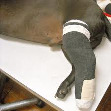
The Schroeder-Thomas splint is another common external splint used to immobilize any fracture below the midfemur of midhumerus. The splint is a traction device or traction splint. This most commonly refers to a splinting device that uses straps attaching over the pelvis, hip, or shoulder as an anchor, metal rods to mimic normal bone stability and limb length, and a sling-like device to apply traction in order to reduce pain and realign the limb. The splint is designed so that bandages and tape are used as slings on the metal frame to position and counteract muscle movement to help align and immobilize parts of the limb.

Although splints are a quick and easy way to set and immobilize fractures, casting is another common form of external coaptation. Casts lie in contact with the skin and are made to conform to the injured area. Plaster and fiberglass can be used for casting, but fiberglass is more commonly used because it is lighter, stronger, and dries quickly. I had the pleasure of seeing a cast applied to a front limb fracture on a dog for the first time the other day. The patient was anesthetized and the fracture was set. Cotton cast padding and a stockingnette were applied to decrease pressure points that could cause skin ulceration. Rolls of casting tape were dunked in warm water and applied up and down the leg. Within minutes the cast was dry and hard as a rock. The cast would be left on for 6-8 weeks in order to allow the fracture to form a callous and heal before being removed.

The Robert Jones bandage is a common external splint used to immobilize lower-limb injuries. This bandage should not be used on upper-limb fractures/injuries because it cannot properly stabilize such injuries. The Robert Jones bandage offers limb stability, tissue fluid absorption, and protects from trauma. It promotes healing by immobilizing the injured area, thus reducing swelling and providing protection from secondary trauma. The bandage consist of multiple layers of soft, compressible, and absorbent materials such as cotton wrap and conforming bandages. These layers are supported by bandage tape stirrups, plastic splints, or a combination of both on either side of the leg. The entire leg is then wrapped with vet wrap and once finished should keep the leg rigid and immobile.
The Schroeder-Thomas splint is another common external splint used to immobilize any fracture below the midfemur of midhumerus. The splint is a traction device or traction splint. This most commonly refers to a splinting device that uses straps attaching over the pelvis, hip, or shoulder as an anchor, metal rods to mimic normal bone stability and limb length, and a sling-like device to apply traction in order to reduce pain and realign the limb. The splint is designed so that bandages and tape are used as slings on the metal frame to position and counteract muscle movement to help align and immobilize parts of the limb.
Although splints are a quick and easy way to set and immobilize fractures, casting is another common form of external coaptation. Casts lie in contact with the skin and are made to conform to the injured area. Plaster and fiberglass can be used for casting, but fiberglass is more commonly used because it is lighter, stronger, and dries quickly. I had the pleasure of seeing a cast applied to a front limb fracture on a dog for the first time the other day. The patient was anesthetized and the fracture was set. Cotton cast padding and a stockingnette were applied to decrease pressure points that could cause skin ulceration. Rolls of casting tape were dunked in warm water and applied up and down the leg. Within minutes the cast was dry and hard as a rock. The cast would be left on for 6-8 weeks in order to allow the fracture to form a callous and heal before being removed.
Thursday, July 11, 2013
Peculiar Poultry
We had the pleasure of treating a rooster at the clinic just the other day. He was a beautiful bird with shiny black feathers and a bright red comb, a show quality bird, but there was one peculiar thing about him. He sat splay legged in his pen with his head tucked under his chest. His neck was so weak and limp that he could barely lift it. The owner explained that it was a perfectly healthy bird and had just started having problems a couple weeks ago, but had gotten progressively worse. The vet examined the rooster and determined that an injury was not the cause of the problem. He concluded that it must be due to a vitamin deficiency. One common deficiency often seen with chickens is riboflavin deficiency. It can be seen rarely in adult birds, but is most commonly seen in young, growing chicks and is often referred to as curled-toe paralysis due to paralysis and disfigurement of the extremities. Sitting splay legged and hock-resting postures are also often seen. The owner was sent home with some riboflavin suspension to give the bird by mouth for 7-10 days, in hopes that the bird would be well and 4-H fair ready within a couple weeks.
Saturday, July 6, 2013
Help! It's stuck!
It seems like it never fails, just when we all think we can close the clinic on time clients walk in at the last minute with animals that need immediate attention and extensive treatment. This always seems to happen on Friday evenings when everyone has plans and is itching to get off work. Just this Friday we had a client call at the last minute to say he was bringing his dog to the clinic because he had a bone stuck in his mouth. We all imagined that the dog had lodged a bone in the back of his mouth and was choking. When the owner arrived with his dog we quickly realized that the dog was in a totally different predicament than we had imagined. The owner had given the dog a stuffed shin bone to chew on. The dog clearly enjoyed his treat a little too much. He had worked so hard to lick the filling from the bone that he somehow worked the bone over his lower jaw and even over his lower canines. The bone was really stuck. The sight was quite hilarious, but the dog was clearly in distress. He clawed frantically at the bone, but it wouldn't budge. The dog was sedated and the veterinarian went to work on trying to remove the bone. He slipped a strand of wire saw underneath the bone and proceeded to saw through. The bone was so tight that the wire saw was starting to cut the dog's skin. The veterinarian went to the clinic's storage room, rummaged around, and came back with a pair of bolt cutters. He placed them at the end of the bone and clamped down. With one cut, the bone snapped off and went flying across the room. The vet laughed and said, "Nothing like an easy and humorous case to end the day."


Sunday, June 30, 2013
Abnormal Eyelids
There were several interesting cases at the clinic this week. One such case involved a young pitbull puppy about five to six months old who had been brought in to be spayed. As I took the dog out of its kennel to be prepped for surgery, I noticed something was not quite right with her eyes. They looked swollen and inflamed and a pus-like discharge was coming from the corners of the eyes. After inspecting her eyes for a few moments I was confident that I knew what the problem was. I had never seen a case in real life, but I remembered reading about the same eye problem in a James Herriot book. I couldn't remember the name of the condition so I asked one of the veterinarians and he told me the dog was suffering from entropion. Entropion is a genetic condition that occurs in a wide variety of dog breeds in which a portion of the eyelid is inverted or folded inward. The inward folding may occur on the top or bottom lid or both. This can cause the eyelashes to scratch and irritate the surface of the eye, resulting in a corneal ulceration. In the long run, entropion can cause a decrease or loss of vision if not treated. Entropion can be treated by using eye drops to alleviate some of the irritation. A temporary surgery known as "tacking" can be performed by rolling the eyelids back and holding them in place with sutures. This surgery is usually performed on young puppies in hopes that the dog will grow into the skin around the eyes. Surprisingly enough, the condition can be permanently corrected by a simple surgery. A small sliver of the affected eyelid or eyelids is removed and the remaining gap is stitched together, which in turn everts the eyelid away from the eyeball.

Sunday, June 23, 2013
The Case of the CoRid Caprine
Although I thoroughly enjoy helping treat dogs and cats of all various shapes, sizes, and breeds, seeing multiples of the same treatment or procedure throughout the week can sometimes become a bit monotonous. It's times like these that I enjoy the rarity of a case involving a species other than a canine or feline. We had such a case at the clinic this weekend involving a caprine, otherwise known as a goat. The owner showed up at the clinic with the goat lying in the back of his truck. It was in very poor shape, thrashing around in the bed and bleating pitifully. The owner explained that he had taken the goat to another clinic where it had been diagnosed and treated for coccidiosis. Coccidiosis is a disease caused by coccidian infection. It is a parasitic disease of the intestinal tract of animals caused by coccidian protozoa. The parasite is passed through fecal-to-oral contact and thrives in overcrowded, dirty, wet pens and unclean water. The disease is very contagious and will quickly spread throughout a herd causing diarrhea (sometimes bloody), dehydration, and fever. The owner confirmed that many other members of his herd had been affected by the disease as well. The owner had been prescribed CoRid (amprolium), a coccidiostat commonly used for the prevention and treatment of coccidiosis. The owner's goats began to improve while using the CoRid, but once again began to decline in health. After hearing this explanation, the veterinarian knew exactly what the problem was. He concluded the goat was suffering from polioencephalomalacia, otherwise known as polio or stargazing. My interest in the case immediately heightened upon hearing this diagnosis. Having learned about polioencephalomalacia in an animal nutrition class this past semester, I was interested in just how a coccidiosis infection had stemmed into polio. The veterinarian explained that although CoRid is commonly used to rid of coccidiosis, if not dosed properly it can cause the problem we were seeing right before us. The drug is a thiamine (vitamin B1) analogue and blocks the thiamine transporter. By blocking thiamine uptake it prevents carbohydrate synthesis, which in turn lowers the supply of carbohydrates to neurons in the brain. The neurons require carbohydrates as an energy source which is necessary for proper nerve function. Without an adequate supply of carbohydrates neurological disorder and neuronal death occur. Thiamine deficiency is one of he main causes of polioencephalomalacia, which is characterized by dullness, depression, convulsions, uncoordinated muscular movements, temporary blindness, abnormal posturing with head thrown backward accompanied by rigidity, and teeth grinding. The goat clearly showed almost all of these signs. The veterinarian felt that the condition had been caught early enough for successful treatment. He administered thiamine intravenously and sent the owner home with instructions to give thiamine injections intramuscularly every 3 to 6 hours.
Thursday, June 20, 2013
ELISA Test
This week the owner and head veterinarian of the clinic took some time to show me how to use the blood analysis machines. The clinic relies heavily on these machines to measure blood cell count, check enzyme and hormone levels, and detect parasites and diseases. These tests are performed routinely throughout the day, and quick results within minutes ensure that clients are provided with conclusive data to back up diagnoses. I found the test kits used along with these machines to be particularly interesting. The clinic uses IDEXX SNAP Tests to screen for many different diseases including, Parvovirus, heartworms, Lyme disease, FIV (Feline Immunodeficiency Virus), and FeLV
(Feline Leukemia Virus). These tests are a simple plastic tray in which a sample, in this case blood, is added to a sample well, the tray is snapped down into place, and within minutes results can be interpreted by the appearance of blue dots in the sample window. It sounds simple enough, but there is actually more to these tests than one might think. Having been exposed to similar types of tests before in class this past semester, I quickly recognized that these SNAP Tests were actually ELISA tests. The ELISA, or enzyme-linked immunosorbent assay, is a test that uses antibodies (proteins produced by the body to identify foreign substances) and color change to detect and identify the presence of a substance, usually an antigen (proteins present on foreign substances like viruses and bacteria that stimulate an immune response).
Here's how the ELISA technology works within these SNAP Tests:
1. A sample and a conjugate are combined in a sample tube. The conjugate contains an antibody with a specific enzyme linked to it. This enzyme antibody complex binds to specific antigens in the sample.
2. The sample tube's contents are poured into the sample well and flow across the surface of the test tray. The conjugate/antigen complex binds to antibody present on the test surface.
3. The device tray is pressed down into place until a snap is heard. The sample flows across the test surface once again in the opposite direction to provide plenty of time for the conjugate/antigen to bind to the antibody.
4. A wash solution clears debris that may interfere with results.
5. A colorless substrate following the wash interacts with the conjugate enzymes. The enzymes convert the substrate molecules from colorless to a blue, amplifying signal. This reaction forms blue dots in the results window for easy interpretation. Each test has a positive control spot that will show, but any color development in the sample spots indicate the presence of specific antigens.
IDEXX SNAP Test with test tray, pipette, blue conjugate, and sample tube
Sunday, June 16, 2013
A Cat Named Dog
As you can imagine there are many interesting and quite hilarious conversations that go on between clients and the assistants/receptionists at the clinic. I thoroughly enjoy listening in on these conversations and found one from this past week to be amusing.
A woman called the clinic to make an appointment for an animal she would be bringing in for treatment. The receptionist proceeded to go through the normal routine of asking for the client's name and contact information. When she asked the client whether she would be bringing in a cat or dog, she replied, "Cat." The receptionist proceeded to ask the name of the cat, to which the client replied, "Dog." The receptionist sat silent for a few seconds and proceeded to ask the name of the dog the client would be bringing in for treatment. The client simply responded, "No, Cat." The receptionist, now thoroughly confused, sat puzzled for a few more seconds before asking, " Ma'am, just what are you bringing, a dog or a cat?" The client replied, "A cat, I am bringing my cat named Dog."

A woman called the clinic to make an appointment for an animal she would be bringing in for treatment. The receptionist proceeded to go through the normal routine of asking for the client's name and contact information. When she asked the client whether she would be bringing in a cat or dog, she replied, "Cat." The receptionist proceeded to ask the name of the cat, to which the client replied, "Dog." The receptionist sat silent for a few seconds and proceeded to ask the name of the dog the client would be bringing in for treatment. The client simply responded, "No, Cat." The receptionist, now thoroughly confused, sat puzzled for a few more seconds before asking, " Ma'am, just what are you bringing, a dog or a cat?" The client replied, "A cat, I am bringing my cat named Dog."
Monday, June 10, 2013
The Importance of Leashes and Pet Carriers
Just this Saturday we had two clients come to the clinic to pick up their cat. The cat had been at the clinic for the past two weeks and had been treated for a blown knee. We were all happy for the cat to finally go home, but as the owners carried it outside it spooked at the sound of the cars going down the highway and ran into the woods in front of the clinic. The owners and I spent a good half hour tromping around in the mud, briars, and poison ivy searching for the cat, but he was nowhere to be found. I told the owners we would keep a look out for the cat and contact them if we found it. I felt sorry for the owners, but I couldn't help but think that such a situation could have been avoided. According to some of the other assistants, a similar event had once occurred when a client came to pick up their dog. The dog was not on a leash and jumped out of its owner's arms. It ended up running out into the road and was hit by a car.
There have been numerous times that I have seen clients bring cats, small dogs, and puppies into the clinic without using leashes or carriers. Just because these animals are small enough to easily carry, it doesn't mean that it is the best way to bring them into public. Some people may think it is cruel to keep their pets restrained or keep them in a carrier, but it can keep you and more importantly your best friend safe. Keeping pets on leashes or in carriers is always a common courtesy to others around and protects both the pets and owners from unexpected accidents. Such forms of animal restraint can prevent animals from chasing or attacking people or other animals. They can also prevent pets from getting away from their owners and fleeing if they become frightened. Proper restraint of animals is a topic that should be stressed more often in animal clinics to ensure the safety of clients and their pets.
There have been numerous times that I have seen clients bring cats, small dogs, and puppies into the clinic without using leashes or carriers. Just because these animals are small enough to easily carry, it doesn't mean that it is the best way to bring them into public. Some people may think it is cruel to keep their pets restrained or keep them in a carrier, but it can keep you and more importantly your best friend safe. Keeping pets on leashes or in carriers is always a common courtesy to others around and protects both the pets and owners from unexpected accidents. Such forms of animal restraint can prevent animals from chasing or attacking people or other animals. They can also prevent pets from getting away from their owners and fleeing if they become frightened. Proper restraint of animals is a topic that should be stressed more often in animal clinics to ensure the safety of clients and their pets.
Saturday, June 8, 2013
Lipoma Lump
This week there were several lump removals at the clinic. I found one of these lump removals to be quite interesting. An owner brought in a Labrador/Mix with a large fatty tumor (about 3.5 inches in diameter) underneath it's left armpit. These tumors are commonly known as lipomas
and are a type of soft tissue tumor. A lipoma is a slow-growing collection of fat cells usually found just underneath the skin. They are soft to the touch, usually movable, and generally painless. Fatty tumors differ from normal fat because they form lumps rather than a flat layer under the skin. Lipomas are a form of cancer, but they are a benign form. This means that the affected cells multiply without normal control, but do not travel throughout the body or invade other tissues. Many lipomas are small in size, but they are capable of enlarging to huge sizes. Usually, treatment of lipomas is not necessary unless they grow very large to the point where they press on internal organs or interfere with walking and movement. Normally these fatty tumors sit in a pocket or fibrous case separated from the surrounding tissues, making it easy for them to be removed by simple excision. Lipomas occur most commonly in older dogs. Some breeds are more likely than others to develop fatty tumors. These include Labrador Retrievers, Doberman Pinschers, and Miniature Schnauzers.

and are a type of soft tissue tumor. A lipoma is a slow-growing collection of fat cells usually found just underneath the skin. They are soft to the touch, usually movable, and generally painless. Fatty tumors differ from normal fat because they form lumps rather than a flat layer under the skin. Lipomas are a form of cancer, but they are a benign form. This means that the affected cells multiply without normal control, but do not travel throughout the body or invade other tissues. Many lipomas are small in size, but they are capable of enlarging to huge sizes. Usually, treatment of lipomas is not necessary unless they grow very large to the point where they press on internal organs or interfere with walking and movement. Normally these fatty tumors sit in a pocket or fibrous case separated from the surrounding tissues, making it easy for them to be removed by simple excision. Lipomas occur most commonly in older dogs. Some breeds are more likely than others to develop fatty tumors. These include Labrador Retrievers, Doberman Pinschers, and Miniature Schnauzers.
Lipoma removed from Labrador/Mix
Sunday, June 2, 2013
The Cost of Prevention: Spaying
There were a wide variety of cases that came into the clinic this week, including pets in need of vaccinations or allergy shots, dermatitis cases, broken limbs, dental cleanings, and even some pets in need of lump removals. The clinic was definitely a blur of cases coming in and out and it would be impossible for me to talk about every one of them, but one case that caught my attention this week was a dog with a mammary tumor.
The mammary gland's function is to produce milk in order to feed offspring. Mammary glands are located in two rows known as the mammary ridges and extend from the chest to the lower abdominal area. The nipples indicate their location on the trunk of the body. A mammary tumor is a benign or malignant tumor of the mammary glands. These benign or malignant tumors occur frequently in unspayed female dogs. Approximately 50% of tumors are found to be malignant and 50% of patients have multiple tumors.
The tumor along with the associated mammary gland was surgically removed from the dog. The surgical process of removing the mammary gland is commonly known as a mastectomy. The tumor was sent off for a histopathology test to determine whether it was benign or malignant. Tumors that are malignant commonly return time and time again and may no longer be able to be removed by surgical means. Chemotherapy may be effective in such cases that indicate lymphatic or vascular invasion, but such treatment may be toxic.
What really troubled me about this case was the fact that it could have easily been prevented/avoided with a simple ovariohysterectomy otherwise commonly known as spay. After doing some research with the help of one of the veterinarians at the clinic I found that females spayed before the first estrous cycle or first heat only had a 0.5% risk for mammary tumors compared to an intact female and those females spayed before the second estrous had only an 8.0% risk of developing mammary tumors. I couldn't understand why people would pass up a simple procedure that could extend the life and wellbeing of their pets, but after some consideration I figured it probably was a matter of cost. I decided to look a little further into the financial aspect of this case. Once again I had one of the veterinarians help me estimate the price breakdown of a general dog spay and a lump removal surgery. The cost breakdown is as follows:
Dog Spay
Canine Spay $91.50
Anesthesia $72.50
Post Surgery Monitoring $0.00
Post Surgery Hospitalization $0.00
Post Surgery Pain Control $20.00
Antibiotic $9.00
Total: $193.00
Lump Removal
Anesthesia $45.00
Lump Removal $95.00
Pain/Inflammation Injection $18.00
Antibiotic $15.00
Histopathology $55.00
Radiograph $86.00
IDEXX Catalyst Chem 17 $68.00
Lasercyte Blood Count $44.00
Total: $426.00
After considering the price breakdown it is obvious that making the decision to spay before the first estrous cycle (around 5-6 months of age) is worth the small investment in the long run. Not only does it save quite a bit of money, it also reduces the risk of cancer in pets allowing them to live a longer, healthier life.
Note: Although this post was addressed toward a case involving a dog, it is important to remember that mammary tumors can commonly occur in cats as well and it is just as important to have them spayed before the first estrous cycle to reduce the risk of cancer.
Thursday, May 30, 2013
Summer Internship
This summer rather than sitting around like most kids and complaining about being bored, I decided to take initiative and get a head start on my career of choice: veterinary medicine. I got ahold of the local veterinary clinic and talked with one of the vets about the possibility of interning for the summer. I laid out my plan of what I wanted to do, topics I was interested in, and what I expected or wanted to learn. So, here I am in my first week of my twelve week journey as a veterinary assistant intern. My daily activities include basic animal care, cleaning cages, kennels, exam tables, and equipment, animal restraint, drawing up shots, preparing animals for surgery, and various other odds and end jobs. Throughout my internship I plan to blog about my experiences and information I have learned. I hope for these posts to be interesting, informative, and beneficial to those who read them.

Subscribe to:
Comments (Atom)